
technology
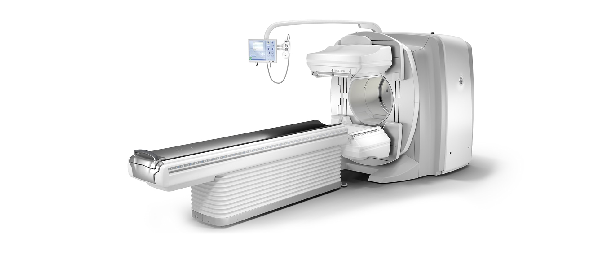
Optimized for everyday imaging
- Provide shorter, more tolerable exams for greater patient comfort with Evolution technology4
- Up to 50 percent reduction in injected dose or scan time with Evolution technology4
- Provide shorter, more tolerable exams for greater patient comfort with Evolution technology4
- Up to 50 percent reduction in injected dose or scan time with Evolution technology4

capability
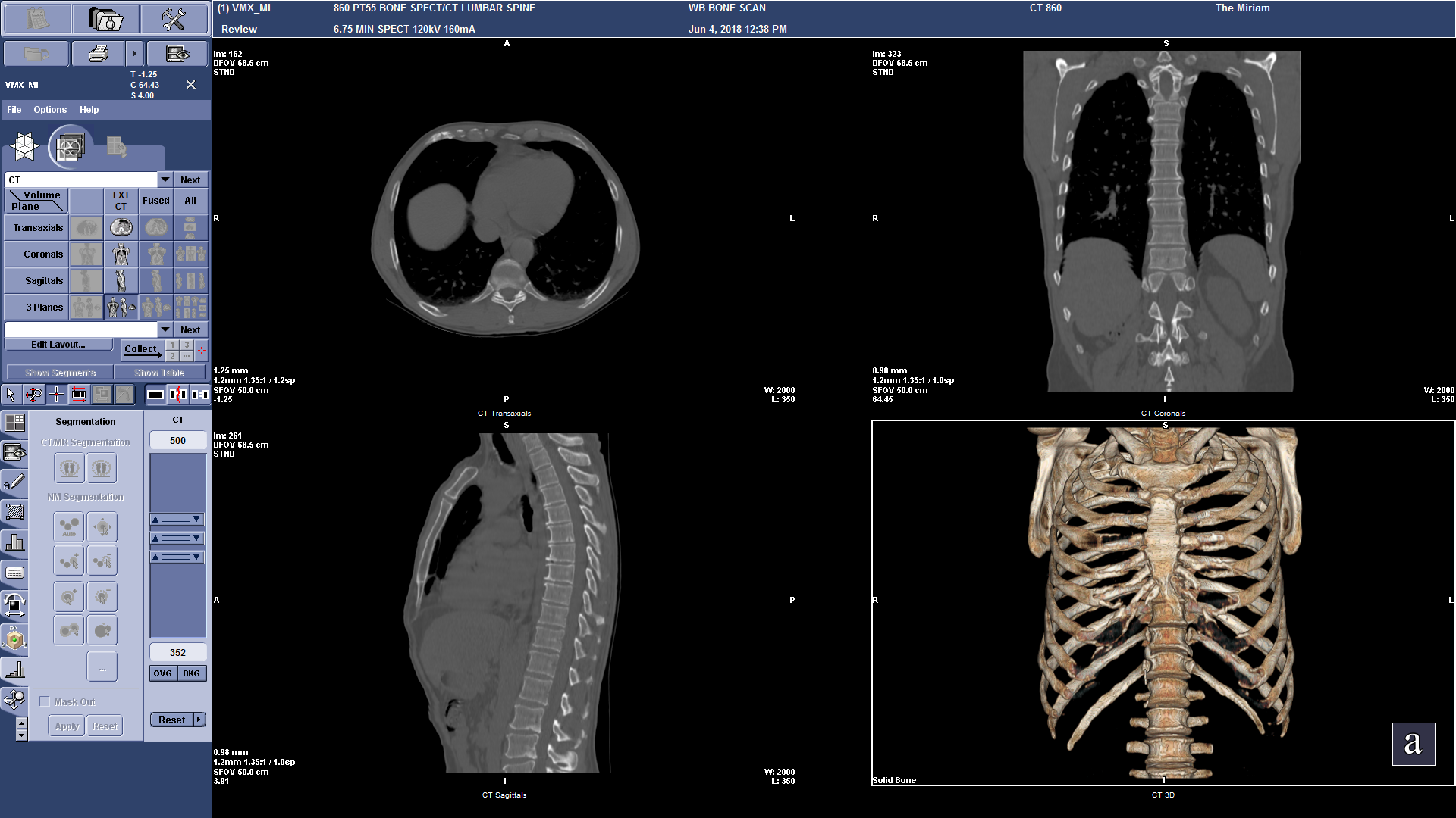
The right mix of SPECT and CT capability
A simpler way to bring everything together
Stay on top of the latest trends in imaging
A CT that stands out when it stands alone

future-ready
A system that’s ready for the future when you are
- Cost-effective access to future innovation
- Access future image quality enhancements and Cloud Analytics with the future-ready SmartConsole
- Reduce cost and save space by sharing collimators with existing 600 Series and 800 Series systems

quantitative applications
Inform your decisions with measurable results
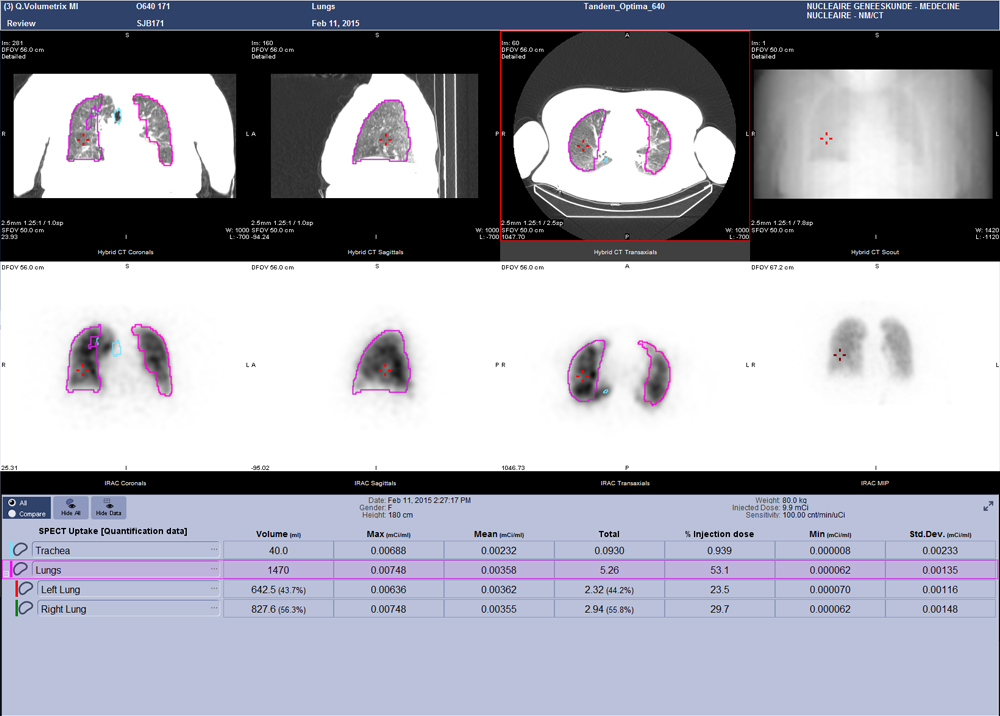
Q. Volumetrix MI
Q. Volumetrix MI
Q. Brain
DaTQUANT™
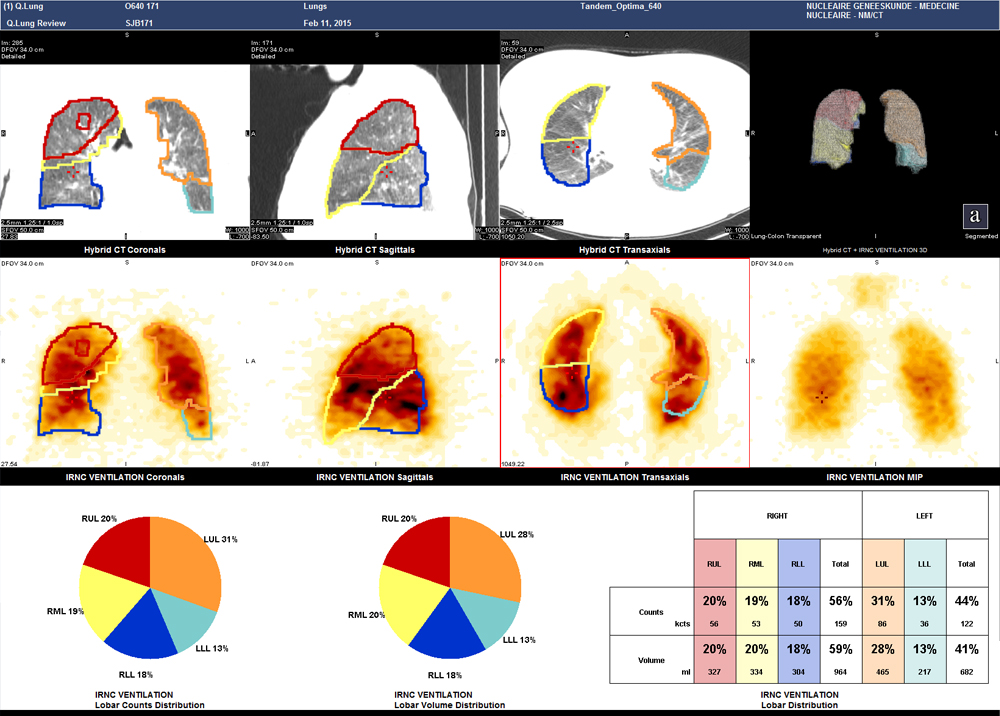
Q. Lung
Q. Lung
Dosimetry Toolkit
summary
A SPECT/CT system for true discovery
Related Products
- 1 Compared to LEHR collimator, with Step & Shoot scan mode (for SPECT) / without Clarity 2D (for Planar). As demonstrated in phantom testing using a bone scan protocol, Evolution processing (for SPECT), and a model observer. Because model observer results may not always match those from a human reader, the actual time/dose reduction depends on the clinical task, patient size, anatomical location and clinical practice. A radiologist should determine the appropriate scan time/dose for the particular clinical task.
- 2 In clinical practice, Evolution options2a (Evolution for Bone, Evolution for Cardiac, Evolution for Bone Planar) and Evolution Toolkit2b are recommended for use following consultation of a Nuclear Medicine physician, physicist and/or application specialist to determine the appropriate dose or scan time reduction to obtain diagnostic image quality for a particular clinical task, depending on the protocol adopted by the clinical site.
- 2a Evolution Options - Evolution claims are supported by simulation of count statistics using default factory protocols and imaging of 99mTc based radiotracers with LEHR collimator on anthropomorphic phantom or realistic NCAT – SIMSET phantom followed by quantitative and qualitative images comparison.
- 2b Evolution Toolkit - Evolution Toolkit claims are supported by simulation of full count statistics using lesion simulation phantom images based on various radiotracers and collimators and by showing that SPECT image quality reconstructed with Evolution Toolkit provide equivalent clinical information but have better signal-to-noise, contrast, and lesion resolution compared to the images reconstructed with FBP / OSEM.
- 3 As demonstrated in phantom testing using a model observer. For SPECT, compared to using the LEHR Collimator and a SPECT Step & Shoot acquisition. For Planar, compared to using LEHR without Clarity 2D.
- 4 In clinical practice, the use of ASiR or VISR may reduce CT patient dose depending on the clinical task, patient size, anatomical location and clinical practice. A consultation with a radiologist and a physicist should be made to determine the appropriate dose to obtain diagnostic image quality for the particular clinical task.